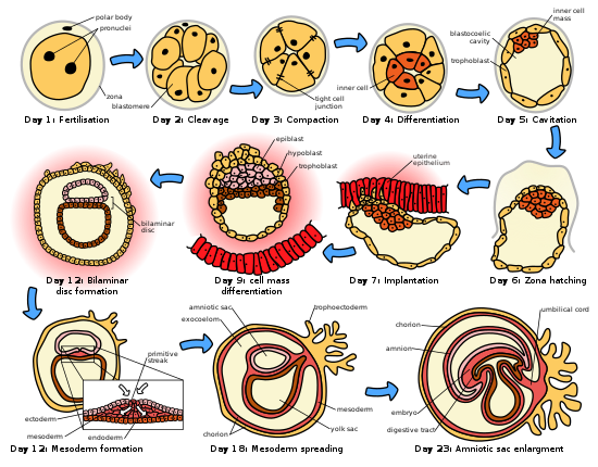
HUMAN EMBRYOGENESIS (STEP 1-4)
Embryogenesis, the first eight weeks of development after fertilization, is an incredibly complicated process. It’s amazing that in eight weeks we’re transforming from a single cell to an organism with a multi-level body plan. The circulatory, excretory, and neurologic systems all begin to develop during this stage. Luckily, like with many complex biological concepts, fertilization can be broken down into smaller, simpler ideas. The big idea of embryogenesis is going from a single cell to a ball of cells to a set of tubes.


Starting from the very beginning
- Step 1: a zygote is the single cell formed when an egg and a sperm cell fuse; the fusion is known as fertilization
- Step 2: the first 12-to 24-hours after a zygote is formed are spent in cleavage – very rapid cell division
The
zygote’s first priority is dividing to make lots of new cells, so
it’s first few days are spent in rapid mitotic division. With each
round of division, it doubles in cell number. This division is taking
place so quickly that the cells don’t have time to grow, so the 32
cell stage known as the morula is
the same size as the zygote. At this point, the zona pellucida (a
protective membrane of glycoproteins that had surrounded the egg
cell) is still intact, which also limits how big it can grow.
Blastulation and Cell Differentiation
- Step 3: during blastulation, the mass of cells forms a hollow ball
- Step 4: cells begin to differentiate, and form cavities
Around day 4, cells continue to divide, but they also begin to differentiate and develop more specific forms and functions. When a cell differentiates, it moves down a certain path toward being a specific type of cell , and this process only goes in one direction. Two layers develop: an outer shell layer known as the trophoblast, and an inner collection of cells called the inner cell mass. Rather than being arranged in a solid sphere of cells, the inner cell mass is pushed off to one side of the sphere formed by the trophoblast. The rest of the fluid-filled cavity is called the blastocoel. The outer trophoblast will develop into structures that help the growing embryo implant in the mother’s uterus. The inner cell mass will continue to differentiate and parts of it will eventually become the embryo This is also the time when the zona pellucida begins to disappear, allowing the ball of cells, now called a blastocyst, to grow and change shape.
At this point, cells in the inner cell mass are pluripotent, meaning they can eventually turn into the cells of any body tissue (muscle, brain, bone, etc). During the second week, these cells differentiate further into the epiblast and the hypoblast, which are the two layers of the bilaminar disc. This disc is a flat slice across the developing sphere, and splits the environment into two cavities. The hypoblast is the layer facing the blastocoel, while the epiblast is on the other side .The
bilaminar disc expands
to fill the space, and becomes the two new cavities: the primitive
yolk sacon
the side of the hypoblast and the amniotic
cavity on
the side of epiblast. The amniotic cavity will eventually surround
the fetus
The hypoblast does not contribute to the embryo, so we will now turn our focus solely on the epiblast. In the next post we will talk about the following phases.


Comentarios
Publicar un comentario